The Role of Imagination and Beauty in Representation: Antibodies as Artistic Objects
Publié par Frédéric Alix, le 31 janvier 2025 1.9k
The Role of Imagination and Beauty in Representation: Antibodies as Artistic Objects
By Frédéric Alix, PhD in Art History
Illustrations in attached file.
On the initiative and with the support of Professors Hervé Watier and Denis Mulleman from the University of Tours. Documents provided by Hervé Watier:
- The representation of antibody reproduction, Paul Ehrlich, On Immunity with Special Reference to Cell Life. Proceedings of the Royal Society of London 66, 424–448 (1900). Figure No. 1.
- Schematic representation of a possible model for gamma immunoglobulin. M. E. Noelken, C. A. Nelson, C. E. Buckley, 3rd, C. Tanford, “Gross Conformation of Rabbit 7 S Gamma-Immunoglobulin and Its Papain-Cleaved Fragments.” J Biol Chem 240, 218–224 (1965). Figure No. 18.
Abstract: For a long time, the Y-shaped silhouette has been firmly established as the visual symbol of the antibody. Simple, direct, and easy to memorize, it is frequently repeated in textbooks and various brochures. It also appears at the heart of many corporate logos. However, although the scientific community is aware that this is merely an outdated cliché, what about non-specialists? This raises the question of what an image conveys within the broader framework of scientific education and public understanding. In this text, we will seek to examine the potential role of creativity in the representation of a scientific object and then illustrate the fact that this representation can be transferred into the potentially more accessible realms of aesthetics and artistic creation.
Introduction
During the COVID-19 pandemic, concepts previously unknown to the general public such as messenger RNA, cytokine storms, or the spike protein entered the mainstream media. At the same time, fanciful and deceitful theories about vaccines were gaining traction. The vast amount of scientific information disseminated, often accompanied by unfamiliar and poorly explained vocabulary, significantly contributed to a sense of confusion among contemporary audiences. The general population, typically armed with only limited knowledge of viruses and the immune system, was confronted with a complex reality that was being challenged by the rise of a “conspiracist” obscurantism fueled by simplistic rhetoric.
“What is rational is real and what is real is rational,” Hegel claimed, yet in this case, distancing ourselves from reality due to a lack of understanding potentially leads us directly into irrationality. And if, as Francisco de Goya warned, “the sleep of reason produces monsters,” then in a hazy space where disconnected signifiers float, where meanings and references are shrouded in a thick veil, a frenzied and militant obscurantism emerges, fed by esoteric and sometimes dangerous discourse.
However, we believe that art has the potential to transform misunderstanding into knowledge. By representing scientific objects such as proteins and antibodies, art can confer upon them the status of artistic objects, transitioning them from the laboratory to the museum, and thereby making them more accessible to the public. By rendering scientific concepts aesthetically pleasing and approachable, art can combat ignorance by stimulating dialogue.
Everyone recognizes the Y-shaped icon meant to represent an antibody. But is this shape truly informative regarding the functions of this particular protein? Art can generate representations that are both scientifically accurate and free from reductive simplifications. To illustrate our argument, we will examine representations created by scientists who had to rely on their imagination and aesthetic sensibility to describe the structure of proteins and antibodies. We will see how these representations moved toward the realm of art. Finally, we will briefly discuss possible artistic interpretations.
A First Image of Antibodies
At the end of the 19th century, the German physician Paul Ehrlich attempted to answer the question of how the body could rapidly produce a large quantity of antibodies when in contact with an infectious agent. He imagined that these antibodies came from certain structures—what we now know as cells—when they were stimulated by a foreign body (antigen); these structures, capable of secreting antibodies, had on their surface a series of antennas (receptors), called "side chains," each capable of interacting with a particular foreign body through chemical bonds. The interaction of a foreign body with a given antenna also triggered the production and secretion of that antenna in the form of soluble antibodies.
Ehrlich turned his theories into images, creating, from what he had imagined, a kind of comic strip narrating the process involved when such an entity comes into contact with a foreign agent[1] (fig. 1). A surprising detail is that Ehrlich believed a single unit could produce antibodies capable of binding to a multitude of antigens. Thus, in these drawings, we see a diversity of shapes meant to represent the so-called "side chains" on the surface of the same structure. Despite the limitations of a hypothesis-based approach, the great advantage of this representation is that it transforms the scientific hypothesis into a visible reality. The ability to translate concepts into drawings was crucial for their dissemination. Ehrlich chose the forms that, in his view, invisible reality might take and did the work of an aesthete by imagining the varied contours of these side chains and antigens.
The first illustration shows the complementarity of side chains with foreign substances. Note how Ehrlich represents a possible fit between the chains and different types of foreign bodies (antigens) through a "key and lock" system. The next image shows a toxin binding to its specific site. In the third image, additional toxins of the same type have attached. The series ends with the fourth illustration: new foreign compounds, identical to the first, bind to the structure, which shows a multiplication of side chains specific to the antigen. Finally, the antibodies detach and enter the bloodstream.
Ehrlich plays with contrast and color alterations and manages to give volume to his representation by balancing peripheral shadows and more central highlights, all in shades of gray. The surface gives the impression of a swelling animated by a granular aspect, like a constellation of craters, offering a sense of relief. But the true stroke of genius lies in the dynamism inherent in this representation as a narrative of a process—a process that includes the "detachment" of antibodies, whose varied spatial arrangements clearly suggest a form of frenetic animation.
Inside the Proteins
About fifty years later, analytical tools and observation instruments had changed, and some of the main issues that Ehrlich had faced were resolved. Knowledge of the structure of antibodies and, more broadly, of proteins increased as accumulated data became significantly more complex. In 1951, the work of chemists Linus Pauling and Robert Corey led to the discovery of the alpha helix[2] (fig. 2) and the beta sheet[3] (fig. 3) as the primary components of the secondary structure of proteins. Pauling understood that hydrogen bonds could maintain the amino acid chain in a helical form and create a three-dimensional structure.
At that time, understanding the structure of atoms and molecules was largely theoretical. Microscopes were incapable of observing molecules, and mastering X-ray diffraction techniques required years of experience. Consequently, Pauling’s theories about chemical bonds were not universally accepted. Sensitive to artistic considerations, he published The Architecture of Molecules in 1964[4], a visually appealing book containing pastel illustrations of chemical bonds drawn by architect and illustrator Roger Hayward (fig. 4). Hayward managed to give volume to each atom by playing with lighting through subtle gradients and the application of white pastel dots. He wrote: “My main skill in my work lies in my interest and ability to think in three dimensions. This comes with a great interest in how things are arranged, both in space and in the physical sense.”
Many years later, Julian Voss-Andreae paid tribute to Pauling with a monumental sculpture (fig. 5). One can clearly see the helical shape, stylized and composed of flat geometric surfaces that extend along the full height.
Three-Dimensional Structures and the Ribbon Model
The second half of the 20th century saw growing efforts to produce a representation capable of integrating advances in research with the need for better understanding protein structure. In 1975, the journal Biochemistry published a representation of the Bence-Jones protein[5]. What stands out is the graphic approach adopting a “ribbon” representation (fig. 6). It was one of the very first representations of this type of alpha-carbon backbone.
One of the two secondary structures, here the beta sheet, is used to highlight the structural properties of the protein as well as the three-dimensional, folded nature of the polypeptide chains. Two elements—elongated bands ending in arrows indicating the direction of the chains, and loops—form the basic vocabulary of this image.
The essential point lies in the clear and concise transmission of information about the structure of this protein, including its V (Variable) and C (Constant) domains. It becomes apparent that this protein unfolds in three dimensions and that pairs of beta sheets, represented by arrowed bands, are interconnected by disulfide bridges, shown here as black rectangles. One also observes that the domains are linked by peptide loops. The numbering corresponds to different amino acid sequences.
The lines are fluid, sometimes broad, and clearly describe the structure. There is no sense of confusion. The twist and overlap of the arrows, depicted as flexible rectangular bands, along with the hatching system, convey an effort to place this figure within a three-dimensional space, creating a sense of depth on a two-dimensional surface. The line seems controlled: it thickens at times to give volume to the sheets and beta strands, which are clearly distinguished.
Such a representation presumably requires a degree of imagination, as it is evident that these shapes cannot correspond exactly to a real protein. In this context, and if the goal is to visualize a concept, a certain level of visual creativity is necessary to make the structure understandable. As with Ehrlich, an aesthetic inventiveness becomes legitimate since it is not essential to faithfully depict the object “in itself” to better understand it.
The Ribbon Model and the Incursion into the Realm of Beauty
In the early 1980s, American biochemist Jane Richardson undertook a very meticulous project of representing protein structures using the ribbon shape, which she refined. Motivated by an artistic sensibility, she experimented with pencil and India ink drawings. Her approach to scientific representation and her interest in aesthetics, combined with her interpretation of the tertiary structure of proteins, eventually led her to create works of art.
This is exemplified by an artistic piece derived from one of the black-and-white models featured in her 1981 article on the anatomy and taxonomy of protein structures[6]. This representation elevates the aestheticization of scientific imagery to an exceptional level (fig. 7). The tertiary structure is exploited purely for its intrinsic visual qualities. Color effects, modulated by light and distributed with great subtlety, enhance the sense of depth across zones and planes. For instance, two shades of green applied to the beta sheets and arrows intensify the perception of a central pocket-like shape, while brown and yellow tones sharply delineate the exterior and interior of alpha helices. The helices are drawn with firm, controlled lines, as are the beta sheets, giving the whole a striking clarity. Iridescent highlights, which add modeling and enhance volume, are visible on the helices. The combination of all these qualities provides remarkable unity to the composition, which almost seems to lift from the background and float on the surface.
Another image shows that, as early as the 1980s, Richardson had complete mastery of various parameters that result in a true artistic creation, offering a kind of classical portrait (fig. 8). This image employs a presentation style borrowing from classical aesthetics, imbued with elegant sobriety. Light effects on shades of gray are skillfully applied, creating modeling that gives volume and movement to the forms. The drawing is flawlessly executed with great fluidity. Although the presentation is “classical,” the object itself—with its twists, asymmetries, and scalloped appearance—recalls the rococo style of 18th-century Europe. The protein’s curves gracefully unfold within a medallion-like frame.
Like Jane Richardson, Byron Rubin has strong ties to Duke University. And, just like Richardson, Rubin dedicated his life to the study of protein structures and also became an artist, creating metal sculptures using the ribbon model to reveal the graceful curvature and flexibility of these structures[7] (fig. 9). The aesthetic qualities of the ribbon model are also found in the work of Mike Tyka. A researcher in protein structures, Tyka focused on protein folding and began making art in 2009. Here we present a sculpture representing the quaternary structure of an antibody protein, specifically an IgG (fig. 10). The work’s title, Savior, is evocative and alludes to the Greek letter gamma (γ) in γ-globulins. Deeply familiar with the subject, Tyka chose to differentiate specific regions of the protein. In the Fab arms of the IgG, with their helices and loops, one can observe the selective application of gold onto the metal, contrasting with the chromed sections. The scientist and artist opted to visually distinguish the antibody’s light and heavy chains.
Some Interpretations
Jane Richardson explained her approach: “Creating these drawings was a fascinating process. On the one hand, the structures are aesthetically very pleasing—particularly, for me, the varied and elegant curves of the beta sheets. On the other hand, making a drawing can alter the scientific understanding of a protein, sometimes revealing a better structural classification or even correcting the trace of a chain.”[8]
The capacity for imagination and creative inspiration was crucial in developing a useful model that allowed researchers to better understand proteins, and consequently, antibodies. Various representations of these scientific objects have illustrated the importance of personal contribution and the freedom of artistic gesture, as shown by Ehrlich, Richardson, and the pioneers of tertiary structure representations.
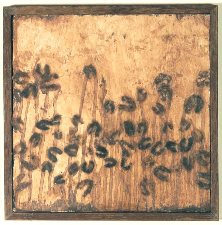
Art can also provide other forms of scientific representation, as seen in the work of Bernard Dublé. His work reflects on the expressive potential of the antibody. Dublé revisits the iconic Y-shape by challenging its rigidity and flatness (fig. 11). The choice of material—a wire—lends itself to multiple twists thanks to its malleability. These twists give volume to the antibody while illustrating its structure of folded polypeptide chains. Furthermore, the antibody’s mobility becomes visible, and the kinetic energy emanating from the object is highlighted (fig. 12). The symbol is in motion, sometimes unfinished, like a non-finito, a kind of raw, dark, and thick sketch with uncertain contours, seeming to glide across a luminous background. The artist multiplies the positions and orientations of the antibody-sign in a dynamic flow that rejects the idea of a static and rigid object.
Other artists, such as Anna Dumitriu, have drawn inspiration from alternative models to the Y-shape, such as the “beaded necklace” (figs. 13[9]–14). Engineered Antibody physically represents the 21 amino acids—plus one added randomly—that make up a modified antibody. This work refers to an experimental practice aimed at deepening the understanding of antibody functionalities, particularly as part of research conducted at the Liu Lab for Synthetic Evolution at the University of California, Irvine[10]. The light and heavy chains of the protein structure were folded in precise accordance with the structure of an antibody.
As for sculpture, we previously mentioned Julian Voss-Andreae, and it would be remiss not to include among his most important and representative works the extraordinary Angel of the West. This sculpture echoes the Vitruvian Man and pays tribute to Leonardo da Vinci (fig. 15). The piece was commissioned by the Scripps Research Institute in Florida, and the antibody appears here as the Vitruvian Man itself[11]. Like Mike Tyka, Voss-Andreae trained in science and has dedicated his art to the structure of proteins.
Beyond the specific domain of antibodies, but still within the realm of proteins, we must highlight the remarkable work of Mara G. Haseltine[12], particularly her piece titled The Waltz of the Polypeptides. This multipart artwork is located on the grounds of the Cold Spring Harbor Laboratory in Long Island, New York. Through this installation, Haseltine illustrates for visitors, students, and faculty the synthesis of a key immune system protein, especially for B cells. From ribosomal synthesis to the completed protein, the different elements of the work—created with a keen eye for visual harmony and staging—integrate seamlessly into the surrounding natural environment (fig. 16).
In a different domain, this time musical and at another level of complexity, artistic creativity can also be used to understand complex processes. For example, consider the interactions between proteins that regulate various biological mechanisms, particularly signaling pathways that trigger immune responses to pathogenic infection. A team of Spanish biochemists has focused on the akirine/subolesin protein, attempting to understand its relationships with the transcription factor NFκB and with proteins RNF10 and THRAPS5[13]. To support this research, a musician collaborated in developing an algorithm to compare the functional interactions of akirine with other proteins across various species. The chosen method involved translating sequences—such as AKR/SUB—into musical scores, thereby creating music based on codons and, consequently, on the amino acids composing these proteins[14] (fig. 17). Could we imagine similar experiments with antibodies? We believe so. Here, the complexity of the artistic approach mirrors that of the scientific research conducted by these Spanish scientists.
To return to something more figurative and directly accessible, artists could also revisit older representations[15] (fig. 18) or create new interpretations, such as those discussed above.
Conclusion
Antibodies, as specific proteins, are complex objects, but as we have seen, it is possible to make this complexity visible in order to better communicate it. Art can help the public gain access to a different level of understanding than what is offered by the Y-shaped representation, enriching scientific culture through a reconnection to tangible and sensory reality. But let us go further: beyond proteins and antibodies, and even beyond biology, why could we not imagine and design artistically relevant representations in other complex fields—representations capable of giving shape to objects whose understanding is often linked to vague or generalized ideas? On this topic, one might think, for example, of the question of atomic orbitals (fig. 19).
To conclude, we would like to give the final word to Jean-Marc Lévy-Leblond, who more than anyone has expressed the existential need to overcome the gap between scientific technicality and sensory reality. Art, we are convinced, is the key to the relationship between reality and science. At the very least, it expresses a scientific reality that it illuminates by stimulating the senses, our sensitivity, by invoking the question of aesthetic Beauty. This is true for antibodies in particular, for proteins in general, and likely for other scientific objects as well.
Jean-Marc Lévy-Leblond wrote: “We scientists need to be reminded of the meaning of immediate reality, not to forget that we no longer work, for a long time now, with the matter of human everyday experience, but with artifacts already highly elaborated by our predecessors. We must rediscover or reestablish the long and tenuous thread that connects theoretical knowledge to sensory curiosity, remember that the cabalistic formulas on our blackboards and the sophisticated devices in our laboratories are closely linked to stones, wind, water, and fire. The words physicists use—even in an old science like mechanics—words such as weight, tension, force, are too easily forgotten to have been borrowed from common language and to have originally designated sensory experiences before becoming the names of elements within an elaborate theoretical framework.”[16]
Art, in our view, is the means to reconnect concept to sensory experience, the development of theoretical knowledge to common language, and is therefore capable of circumscribing the “sleep of reason.”
---
[1] P. Ehrlich, On immunity, with particular reference to cellular life. Proceedings of the Royal Society of London 66, 424-448. (1900).
[2] L. Pauling, R. B. Corey, H. R. Branson, “The structure of proteins; two hydrogen-bonded helical configurations of the polypeptide chain”. Proc Natl Acad Sci U S A 37, 205-211 (1951).
[3] L. Pauling, R. B. Corey, “Configurations of Polypeptide Chains with Favored Orientations Around Single Bonds: Two New Pleated Sheets”. Proc Natl Acad Sci U S A 37, 729-740 (1951).
[4] L. Pauling, Roger Hayward, The architecture of molecules, W. H. Freeman and company, San Francisco, London, 1964.
[5] A. B. Edmundson, K. R. Ely, E. E. Abola, M. Schiffer, N. Panagiotopoulos, “Rotational allomerism and divergent evolution of domains in immunoglobulin light chains”. Biochemistry 14, 3953–3961. (1975).
[6] S. Richardson, “The anatomy and taxonomy of protein structure”. Adv Protein Chem 34, 167-339 (1981).
[7] Personal website : http://molecularsculpture.com/
[8] J.S. Richardson, “Early ribbon drawings of proteins”. Nat Struct Biol 7, 624-625 (2000).
[9] IMGT®.
[10] According to Professor Xiang Li: “Working with Anna on the antibody necklace piece actually made me realize that I had an error in the sequence of my antibody that I am using in my research project. To build the work we had to compare my antibody sequence to the correct antibody sequence in a crystal structure, and I noticed that those sequences did not match. Since then, I have fixed the sequence of my antibody for my research project!”.
[11] To see more artworks representing proteins created by Julian Voss-Andreae: https://julianvossandreae.com/works/protein-sculptures-outdoor-works/
[12] Personal website: https://www.calamara.com/artwork/waltz-of-the-polypeptides/
[13] Artigas-Jerónimo S, Pastor Comín JJ, Villar M, Contreras M, Alberdi P, León Viera I, Soto L, Cordero R, Valdés JJ, Cabezas-Cruz A, et al. A Novel Combined Scientific and Artistic Approach for the Advanced Characterization of Interactomes: The Akirin/Subolesin Model. Vaccines. 2020 ; 8(1):77. https://doi.org/10.3390/vaccines8010077
[14] To listen an extract: https://freesound.org/people/josedelafuente/sounds/478998/
[15] M. E. Noelken, C. A. Nelson, C. E. Buckley, 3rd, C. Tanford, “Gross Conformation of Rabbit 7 S Gamma-Immunoglobulin and Its Papain-Cleaved Fragments”. J Biol Chem 240, 218-224 (1965).
[16] J.M Lévy Leblond, La pierre de touche, Paris, Gallimard, 1996, p. 174-175, quoted in Jean-Paul Charrier, Scientisme et Occident, Essais d’épistémologie critique, Paris, Connaissances et Savoirs, 2005, p.312-313.